Categories
Uncategorized

The Role of Interventional Neuroradiology in Treating Cerebral Arteriovenous Malformations (AVMs)

Arteriovenous malformations (AVMs) are abnormal tangles of blood vessels that can occur in various parts of the body, including the brain. These complex vascular anomalies pose significant health risks and require specialized treatment approaches. In recent years, interventional neuroradiology has emerged as a ground-breaking field, offering minimally invasive techniques for diagnosing and treating AVMs with remarkable precision and effectiveness. This blog explores the role of interventional neuroradiology in managing arteriovenous malformations, highlighting its benefits and advancements.

Understanding Arteriovenous Malformations:

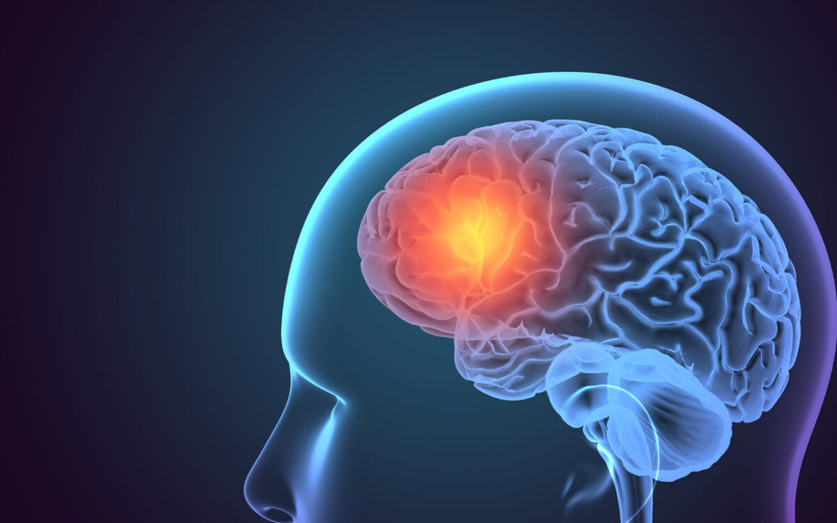
Arteriovenous malformations involve an abnormal connection between arteries and veins, bypassing the capillary network and normal tissue. These tangled vessels disrupt normal blood flow, increasing the risk of bleeding, ischemia, and other complications. Traditional treatment methods, such as open surgery, carried significant risks and had lengthy recovery periods. However, interventional neuroradiology has revolutionized the management of AVMs through innovative procedures that minimize invasiveness and maximize patient outcomes.

Embolization Therapy:

One of the primary interventional neuroradiology techniques used in AVM treatment is embolization therapy. This procedure involves the use of catheters to deliver embolic agents directly into the abnormal blood vessels. The embolic agents cause the vessels to close off, reducing blood flow within the AVM. Embolization can be used as a stand-alone treatment or as a preoperative measure to reduce the size and vascularity of the malformation before surgical intervention.

Radiosurgery:

Radiosurgery involves delivering a precisely targeted dose of radiation to the AVM, causing the blood vessels to gradually close off over time. This technique, performed using advanced imaging guidance, offers a non-invasive alternative to traditional surgical interventions. It allows for the precise delivery of radiation to the AVM while minimizing damage to surrounding healthy tissues.

Advantages of Interventional Neuroradiology:

The utilization of interventional neuroradiology techniques in AVM treatment offers several advantages. First and foremost, these procedures are minimally invasive, resulting in shorter hospital stays, reduced risk of complications, and faster recovery times for patients. Additionally, interventional neuroradiology allows for greater precision in targeting abnormal blood vessels, ensuring optimal outcomes. The collaborative approach between interventional neuroradiologists and neurosurgeons also enables a multidisciplinary treatment strategy that maximizes patient care.

Conclusion:

Interventional neuroradiology has transformed the treatment landscape for arteriovenous malformations, providing effective alternatives to traditional surgical approaches. Through embolization therapy and radiosurgery, interventional neuroradiologists can precisely target and treat AVMs with minimal invasiveness and improved patient outcomes. By leveraging innovative imaging techniques and catheter-based procedures, this field continues to advance, offering hope and relief to individuals affected by these complex vascular anomalies.

Discover the cutting-edge world of interventional neuroradiology and its role in revolutionizing AVM treatment. Explore our comprehensive range of services for effective management and improved patient care. Contact us today for a personalized consultation.

Categories
Uncategorized

Headache or Stroke Symptoms? It Could Be a Cerebral Aneurysm – Know the Warning Signs

A Headache can be a sign of many things, from stress to dehydration, and usually, it’s nothing to worry about. However, in some cases, a headache can be a warning sign of something more serious. One of those things is a cerebral aneurysm, which is a bulge in a blood vessel in the brain that can rupture and cause a potentially life-threatening condition known as a subarachnoid hemorrhage.

A cerebral aneurysm can be present for years without causing any symptoms. However, when an aneurysm ruptures, it can cause a sudden and severe headache, often described as the “worst headache of your life.” This headache is usually accompanied by other symptoms, such as nausea, vomiting, sensitivity to light, and a stiff neck. In some cases, the ruptured aneurysm can also cause stroke-like symptoms, such as weakness or numbness on one side of the body or difficulty speaking.

If you experience any of these symptoms, it’s crucial to seek medical attention immediately. A ruptured cerebral aneurysm is a medical emergency that requires prompt treatment to prevent permanent brain damage or even death. Don’t wait, call Dr. Vivek Gupta today for expert advice and care.

What is a Cerebral Aneurysm?

A cerebral aneurysm is a bulge in a blood vessel in the brain. It can be present for years without causing any symptoms, and many people don’t even know they have one. However, when an aneurysm ruptures, it can cause a sudden and severe headache, often described as the “worst headache of your life.” This headache is usually accompanied by other symptoms, such as nausea, vomiting, sensitivity to light, and a stiff neck. In some cases, the ruptured aneurysm can also cause stroke-like symptoms, such as weakness or numbness on one side of the body or difficulty speaking.

What are the Warning Signs of a Cerebral Aneurysm?

The warning signs of a cerebral aneurysm can vary depending on the size and location of the aneurysm. However, some common symptoms include:

Severe Headache

This is the most common symptom of a ruptured aneurysm. It’s usually described as a sudden, intense headache that is different from any headache you’ve had before. It can be accompanied by a feeling of pressure in the head, ringing in the ears, and a stiff neck.

Neurological Symptoms

These can include difficulty speaking, weakness or numbness on one side of the body, vision changes, and loss of coordination. In some cases, the neurological symptoms can be the only symptoms of a cerebral aneurysm.

Other Symptoms

Other symptoms of a ruptured aneurysm can include nausea and vomiting, sensitivity to light, and a stiff neck. These symptoms can develop suddenly, and they require immediate medical attention.

Why is it a Medical Emergency?

A ruptured cerebral aneurysm is a medical emergency that requires prompt treatment to prevent permanent brain damage or even death. The bleeding from the ruptured aneurysm can cause a stroke or lead to other complications, such as hydrocephalus (an accumulation of fluid in the brain) or vasospasm (a narrowing of the blood vessels in the brain). Prompt diagnosis and treatment can help reduce the risk of complications and improve the chances of a full recovery.

If you experience any of the warning signs of a cerebral aneurysm, seek medical attention immediately. Contact us today to learn more about brain health and how to reduce your risk of developing a cerebral aneurysm.

Categories
Uncategorized

How High Blood Pressure Can Lead to Stroke

High blood pressure, also known as hypertension, is a common health condition that affects millions of people worldwide. It occurs when the force of blood against the walls of your arteries is too high, which can damage the blood vessels and increase the risk of serious health problems, including stroke. In this blog, we’ll explore how high blood pressure can lead to stroke and what steps you can take to reduce your risk.

What is a stroke?

A stroke occurs when the blood supply to your brain is disrupted, either by a blood clot or a burst blood vessel. When this happens, brain cells begin to die, which can lead to permanent brain damage, disability, and even death. There are two main types of strokes: ischemic stroke, which is caused by a blood clot, and hemorrhagic stroke, which is caused by a burst blood vessel.

How does high blood pressure increase the risk of stroke?

High blood pressure can weaken the walls of your blood vessels, making them more prone to bursting. When a blood vessel in your brain bursts, it can cause a hemorrhagic stroke, which can be life-threatening.

If you have high blood pressure, there are several steps you can take to reduce your risk of stroke:

Manage your blood pressure: Work with your healthcare provider to manage your blood pressure by following a healthy diet, exercising regularly, and taking medications as prescribed.

Maintain a healthy weight: Being overweight or obese can increase your risk of high blood pressure, so it’s important to maintain a healthy weight through diet and exercise.

Exercise regularly: Regular exercise can help lower your blood pressure and reduce your risk of stroke. Aim for at least 30 minutes of moderate-intensity exercise most days of the week.

Eat a healthy diet: A diet that is rich in fruits, vegetables, whole grains, and lean protein can help lower your blood pressure and reduce your risk of stroke.

Quit smoking: Smoking can damage your blood vessels and increase your risk of stroke, so it’s important to quit smoking if you’re a smoker.

Manage stress: Chronic stress can increase your blood pressure, so it’s important to find healthy ways to manage stress, such as meditation, yoga, or deep breathing exercises.

Follow your healthcare provider’s recommendations: If you have other medical conditions that increase your risk of stroke, such as diabetes or heart disease, it’s important to follow your healthcare provider’s recommendations for managing these conditions.

In conclusion, high blood pressure is a major risk factor for stroke, but there are steps you can take to reduce your risk. By managing your blood pressure, maintaining a healthy weight, exercising regularly, eating a healthy diet, quitting smoking, managing stress, and following your healthcare provider’s recommendations, you can reduce your risk of stroke and protect your brain health.

“Take control of your heart health and reduce your risk of stroke by making healthy lifestyle choices. Contact us today to learn more about stroke prevention and blood pressure control.

Categories
Uncategorized

Faces Of Stroke

Picking up a stroke early aids in preventing brain damage and complications associated with the condition. Looking at a person’s face can help you understand if they are getting a stroke.

So, how does a face of a stroke patient look like? Read on to know more about it.

A Word About Stroke

A stroke is a condition where a blood vessel supplying to the brain ruptures or is blocked by a blood clot. In either case, a part of the brain cannot receive the nutrients and oxygen required for its survival. And if the blood supply is not retrieved, that part of the brain may be damaged, resulting in various complications.

It is thus crucial to identify a stroke early and treat it immediately. One of the common symptoms can be understood by looking at the person’s face.

What To Look For On The Face?

Symptoms of stroke appear on the part of the body supplied by the damaged brain areas.

F.A.S.T. is an easy way to identify the immediate signs of a stroke.

Face: The face droops on one side or feels numb. You can ask the person to smile. The smile of someone with a stroke is not symmetrical, and lips droop on one side of the face.

In addition to the face, other signs include:

Arm Weakness: The arm may feel numb and/or weak. Ask the person to raise both arms. The affected arm drifts downwards.

Speech Difficulty: The person may find it difficult to speak or have slurred speech. You can also tell them to speak a sentence such as “the sky is blue.” A person with a stroke will find it challenging to say the sentence clearly.

T in the acronym indicates timely intervention. If you feel that someone has a stroke, seek medical care immediately.

Other symptoms of a stroke are:

  • Confusion or lack of responsiveness
  • Vision problems such as double or blurred vision
  • Loss of balance and trouble walking
  • A sudden, severe headache
  • Dizziness
  • Seizures
  • Vomiting or nausea
  • Numbness and weakness in one side of the body

Recovery

Once a stroke is managed, the person can recover within days, weeks, or months, depending on their condition. They can return to their normal activities without needing external aid in most cases. Medicines and physiotherapy aid in faster recovery from a stroke.

Categories
Uncategorized

Arterial Ischemic Stroke in Children

Arterial ischemic stroke is a rare but crucial cause of complications in children. Early diagnosis and treatment aid in preventing complications.

Arterial ischemic stroke

An arterial ischemic stroke is an injury in the spine or brain because of impaired oxygen and nutrient supply to the affected area. In most cases, ischemic stroke is caused by damaged or narrowed arteries or obstruction due to blood clots.

Fortunately, children recover quickly and completely as compared to adults. The reason is that young blood vessels and brains adapt quickly after an injury.

While some strokes have none or minimal effects, others can cause complications depending on the part of the brain affected, the severity of the stroke, and how early the treatment was initiated.

Complications may include sensory loss, weakness, language impairment, or visual changes.

Symptoms In Children

Symptoms of arterial ischemic stroke usually appear suddenly and affect one side of the child’s body. Newborns have no symptoms. Infants may have unusual irritability or seizures.

Symptoms in older children include:

  • Numbness or weakness on one side of the body
  • Seizures affecting one side of the body
  • Difficulty in speaking
  • Loss of balance or dizziness
  • Trouble walking
  • Vision problems
  • A sudden severe headache associated with double vision, sleepiness, or vomiting

If your child has any of the mentioned symptoms, seek medical care immediately.

Causes

Two leading causes for obstructed blood vessels in children include:

Blood clots because of:

  • Blood diseases such as clotting factors
  • Severe infections
  • Congenital heart conditions
  • Abnormal blood clotting

Damage to or abnormalities of the arteries because of:

  • Injury to the head or neck
  • Inflammation of blood vessels
  • A tear along lining of a blood vessel

Management

The management of stroke in children is different from adults. A child with a stroke generally receives blood thinning drugs. They help in clearing the clot and improve the blood flow. The clot may be removed using catheter-based devices in certain cases (Uncommon). For inflamed blood vessels causing a stroke, medicines to soothe inflammation are required.

Once the blood flow is retrieved, the child may need some care, including physiotherapy exercises, to have a better quality of life.

Categories
Uncategorized

Head, Neck, and Brain Tumor Embolization

What is Head, Neck, and Brain Tumor

Head, neck, and brain tumors are often very complex and may require a combination of special approaches in resolving them. Embolization is an integral part of managing these tumors.

Embolization refers to a procedure that can block blood flow to an area of the body. The brain or spinal cord tumors can be challenging to remove or may not be accessible for removal.

Reducing or cutting the blood supply to such tumors before the surgery can help to improve the effectiveness and lessen the complications. Thus, it has become adjuvant to the surgical treatment of these tumors.

The endovascular approach is the most common type of widely used approach. It is an invasive surgical procedure used to block blood vessels supplying the tumor thereby making the tumor removal safer and with less blood loss.

Embolization In Treatment Of Tumors

The procedure involves making a tiny incision in your groin area, and a catheter is inserted via a blood vessel (femoral artery). The catheter is guided through the body using X-rays. When the catheter reaches the site that needs to be treated, the material is injected to seal your blood vessel (metal coils, PVA particles, gelfoam, liquid embolics). The material used will be decided by your interventionist.

Embolization of head, neck, and brain tumors may be recommended to treat the cancer growth in the following cases:

  • Carotid body tumors
  • juvenile nasopharyngeal angiofibroma (JNA)
  • Larynx Meningiomas (brain and spine)
  • Cerebral Hemangiopericytomas
  • Paragangliomas: in neck, skull base or ear

Advantages of Embolization

The advantage of embolization includes:

  • To reduce tumor recurrence
  • To control surgically inaccessible arterial sites
  • To allow better visualization of the surgical field and decreased surgical complications
  • To relieve uncontrollable pain
  • To reduce the blood loss during the surgery
  • To reduce the risk of damage to nearby tissues
  • Shorten the operative time and less invasive
  • To increase the chances of complete surgical resection

Preparation

Endovascular embolization is performed in a hospital by an interventional neuroradiologist. It is often performed as an elective preoperative procedure. The following things should be kept in mind:

  • Inform your surgeon about all medications that you take, including prescription, non-prescription, vitamins, and herbal supplements
  • If you smoke or take alcohol regularly
  • If you are having fever.
  • If you are on any blood thinning agents.

Recovery

Depending upon your recovery, you will be asked to stay in the hospital for 1 or 2 days. You might be asked to stay longer if there were any complications during the procedure. Usually, your rate of recovery will determine your discharge. Your comorbidities may also affect your recovery speed. The area where the incision was made may remain sore for several days.

Your physician will ask you to follow up depending on the disease and closely monitor you.

Categories
Uncategorized

Can We Prevent A Second Stroke?

To answer the question mentioned above, it is first essential to understand the basics of Stroke.

Keep reading the article to understand and find out if we can prevent second stoke and how we can prevent it?

What is Stroke?

A stroke is a condition that occurs when a blood vessel in the brain bursts and bleeds or when there is a blockage of the blood supply to the brain. Both scenarios (rupture or blockage) prevent blood and oxygen from reaching the brain’s tissues.

Without the supply of oxygen, brain cells and tissues become damaged and begin to die immediately. As the brain cells die, people may experience weakness or paralysis and lose walking or speaking.

There are three main types of strokes they are classified as follows:

  • Ischemic Stroke: This is the most common type of stoke, and it results because of a blood clot that prevents blood and oxygen from reaching an area of the brain
  • Hemorrhagic stroke: This occurs when a blood vessel ruptures. The rupture is mainly due to aneurysms (formation of a balloon and weakening in a part of the artery)
  • Transient ischemic attack: It occurs when blood flow to a part of the part is inadequate for a brief amount of time. Normal blood flow resumes after a short period, and the symptoms during the episode resolve without any treatment. It is known as a ministroke.

Strokes are fatal. The article explains if a person can prevent a second stroke and how it can be prevented.

Can we prevent a second Stroke?

Yes, it is possible to prevent a second stroke. The causes for the second stroke are majorly similar to a primary stroke.

Researchers believe that you are most vulnerable for three months to 1 year after your first stroke. So even after surviving one stroke, you may still need to take a lot of care to prevent the second stroke.

Causes of the second stroke

The causes of one or more strokes mainly remain the as which are as follows:

  • Diabetes: High blood sugar levels damage the blood vessels, increasing the possibility of clot formation in the blood vessel.
  • High blood pressure: Untreated or uncontrolled blood pressure doubles the risk of stroke.
  • High cholesterol: High cholesterol levels in your blood can lead to plaque formation. Thus, it causes a decrease in blood flow to the brain and other parts of the body.
  • Smoking: Smoking causes thickening of the blood and increases plaque formation in the arteries, which eventually may decrease blood flow to the brain or other parts of the body.
  • Obesity: Being overweight increases the odds of stroke and is linked to other diseases (heart diseases, diabetes, and high blood pressure).

Prevention

Some tips may help to prevent a second stroke:

  • Taking medication: If your physician has prescribed medications to control cholesterol, diabetes, or blood pressure, make sure you take medications regularly. You should not miss your doses. Even if you feel better, you should not discontinue your medications. Not taking medication is a significant risk factor that may cause stroke again.
  • Exercise: You should work out (cardio) for at least two and half hours a week. Aerobic exercises such as walking and riding a bicycle can help you manage the risk factors of stroke.
  • Healthy lifestyle: You should incorporate healthy eating habits. Avoid junk food and shift to the Mediterranean (fruits and vegetables, whole grains, and low processed foods). Avoid or restrict salt consumption. You should quit smoking and use aids like nicotine patches if you are smoking. It is also best to avoid or quit alcohol consumption.

These changes help reduce the chances of stroke and help manage other diseases such as obesity, diabetes, blood pressure, or high cholesterol.

Categories
Uncategorized

What Is the Relevance of Time in Acute Stroke Treatment?

A stroke is a condition where the blood supply to the part of the brain is reduced or blocked, affecting its oxygen and nutrient supply. The main cause is a blood clot in the artery supplying the brain (ischemic stroke).

Without oxygen and nutrients, brain cells begin to die. Hence, stroke is a medical emergency that requires prompt treatment. Early action can significantly reduce damage to brain cells and other complications.

Let’s understand how timely intervention helps anyone with stroke.

Symptoms Of Stroke

To get timely treatment, it’s essential to understand what stroke looks like. While it may not always be easy to pick up stroke, look out for the following symptoms:

  • Face: One side of the face drops while smiling
  • Arms: Try to raise both arms. You may find it difficult to raise one arm, or the arm drifts downwards.
  • Speech: You may have strange or slurred speech
  • Time: If you or anyone has the above signs, it’s recommended to seek emergency medical care.

Relevance Of Time in Acute Stroke Management

According to the American Stroke Association and American Heart Association: “Time lost is brain lost.

Once you reach the hospital, the doctor will ask for your medical history and when you noticed them. They will also run brain scans to understand the type of stroke you have.

As discussed above, the earlier the treatment, the lesser the complications.

Do what does early mean?

Research shows that seeking medical treatment within 3 to 4.5 hours of an acute ischemic has a better outcome. Medical intervention in the mentioned time frame allows the doctor to treat you with a type of clot-busting medicine, known as Tissue plasminogen activator (tPA)

Getting tPA significantly improves your chances of recovering from a stroke. Studies have shown that people getting tPA have a lesser disability than people not getting tPA.  Patients who receive timely tPA have lesser need of long-term care in the nursing home.

The newer treatment Mechanical Thrombectomy can be done even upto 24 hours but not in all patients. It is an excellent technique to treat stroke especially if large artery is blocked. However like tPA earlier the Thrombectomy is done better is the clinical outcome of stroke patients.

Categories
Uncategorized

Cerebral Aneurysm Coiling

Cerebral aneurysm is an abnormal dilation or bulge or ballooning of an artery in the brain, mainly occurring where the artery divides. Weakening of the muscular layer of the artery makes it susceptible for this abnormal dilation. It may leak or rupture, which causes bleeding into the brain, resulting in hemorrhagic stroke. A ruptured aneurysm is often life-threatening and requires immediate medical attention.

What is Cerebral aneurysm coiling?

Cerebral coiling or endovascular coiling is a minimally invasive procedure that does not require an incision on the skull. However, it is carried out under general anesthesia. The procedure is performed with the aim to block the blood flow into the aneurysm. A catheter is placed into the artery through your groin or wrist in this technique. That catheter is guided into the brain and into the aneurysm. After reaching the aneurysm, a thin platinum coil is inserted into the aneurysm and detached. Multiple such coils are packed inside the aneurysm, blocking the blood flowing inside the aneurysm, leading to sealing off from the main artery. Over a period, a clot is formed inside the aneurysm, which prevents it from growing or rupturing.The coils remain permanently inside the aneurysm. They are safe and long term.

Why is Cerebral aneurysm coiling done?

It is an effective treatment for patients having ruptured or unruptured aneurysms. However, it is always done if the aneurysm has already ruptured. Case where bleeding has not occurred the coiling is done after assessing the bleeding risk which depends upon many factors like size/configuration of the aneurysm, age, co-morbidities, medications history etc.

Risks associated with Cerebral aneurysm coiling

Though the procedure is minimally invasive, it may cause some side effects. The complications associated with the procedure are infection, stroke, seizure, allergic reactions, and bleeding.

Post-surgery care

After the surgery, you may be shifted to the intensive care unit for further observation and monitoring. If the coiling is done for unruptured aneurysm the hospital stay might be of 3-4 days. If it is after bleeding has occurred then the hospital stay may extend to 10-14 days. The patient may feel mild tenderness in the groin or leg region. Your physician will provide you with specific detailed instructions regarding the post-surgery.

The following set of restrictions are be expected to be given by the surgeon:

  • Do not consume alcohol as it causes thinning of blood, and you will be at risk of bleeding
  • Do not smoke or use nicotine products
  • Do not lift heavy objects or perform strenuous activities for the next three days
  • Do not drive unless your surgeon says
  • Drink plenty of water to flush out the contrast dye
  • Take the prescribed medications on a timely basis
Categories
Uncategorized

Intracranial aneurysm

Intracranial aneurysms are known by various names such as brain aneurysms, cerebral aneurysms, or berry aneurysms or sometimes called ballooning of the brain blood vessels. Most types of brain aneurysms do not show any symptoms. They start showing symptoms when they become large, burst, or begin to leak blood.
To understand more about it, keep reading.

What is an Intracranial aneurysm?

An intracranial aneurysm is an abnormal dilation or bulge or ballooning of an artery in the brain. It mainly occurs due to the weakening of the muscular layer of the artery. The image on a scan often resembles a berry hanging on a stem; hence it is known as a berry aneurysm.
It may leak or rupture, which causes bleeding into the brain, resulting in hemorrhagic stroke. A ruptured aneurysm may be critical and requires immediate medical attention.

Symptoms

Severe headache is an important symptom in an aneurysm. These headaches often are sudden and are deemed as the worst headache ever experienced. Other symptoms include:

  • Stiff neck
  • Nausea
  • Vomiting
  • Double or blurred vision or change in vision
  • Confusion
  • Drooping eyelid
  • Loss of consciousness
  • Sensitivity to light
  • Pain above or behind the eye
  • Weakness on one side of the body or face
  • Dilated pupils

Causes

There are various risk factors, which may contribute to the formation of aneurysms. They are listed as follows:

  • Aging
  • Genetics (history of intracranial aneurysm in the family)
  • Smoking
  • High blood pressure
  • Severe head injury or trauma
  • Use of cocaine or other drugs
  • Congenital abnormality
  • Infection
  • Excessive alcohol consumption
  • Women are at a higher risk
  • Other disorders such as Ehlers-danlos syndrome, fibromuscular dysplasia, Marfan syndrome, and autosomal dominant polycystic kidney disease

Diagnosis

Family history is taken at the beginning to understand the history of a brain aneurysm in the family. If you are experiencing any of the above symptoms, your physician may ask you to undergo any of the following screening tests:

  • CT scan of the brain
  • MRI of the brain
  • Cerebrospinal fluid test
  • Cerebral angiogram (DSA)

Treatment

The ideal treatment option depends on various factors such as size, location of the aneurysm, medication condition of the patient, availability of other treatment options, and age of the patient. The goal of the treatment involves preventing the aneurysm from bleeding or bleeding again. The following are some of the commonly available treatment options:

  • Endovascular Coiling treatment: In this treatment, the surgeon inserts a catheter (a hollow plastic tube) into the blood vessel (artery) and threads it through your body to the affected region. The entire treatment is then done through this tube/catheter by packing the aneurysm with coils from within.
  • Surgical clipping treatment: In this procedure, the surgeon identifies the aneurysm and the artery that is affected. The procedure involves removing a skull section to enter the affected region. The neurosurgeon places a tiny clip on the affected artery to stop the blood flow.
  • Flow diverters treatment: This newer treatment option diverts the blood flow away from the affected region. The diversion created restricts the blood movement within the affected area and promotes healing.

Pt. will require additional therapies like:

  • Rehabilitative therapy: This treatment option is opted when damage to the brain occurs from a subarachnoid hemorrhage that affects speech, physical, sensory, or cognitive problems.
  • Medications: Various medications such as calcium channel blockers, pain killers, anti-epileptic medications may be prescribed.