Headaches are one of the most common health complaints and are often linked to stress, dehydration, migraines, or lack of sleep. However, in some situations, a sudden or unusually severe headache may signal a more serious neurological condition such as a brain aneurysm. According to Dr. Vivek Gupta, understanding the warning signs of brain aneurysm and knowing when to seek medical attention can play an important role in protecting brain health and preventing complications.
What Is a Brain Aneurysm?
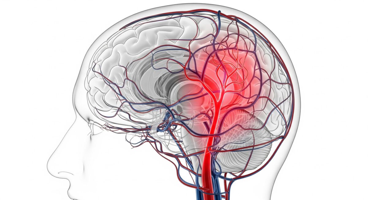
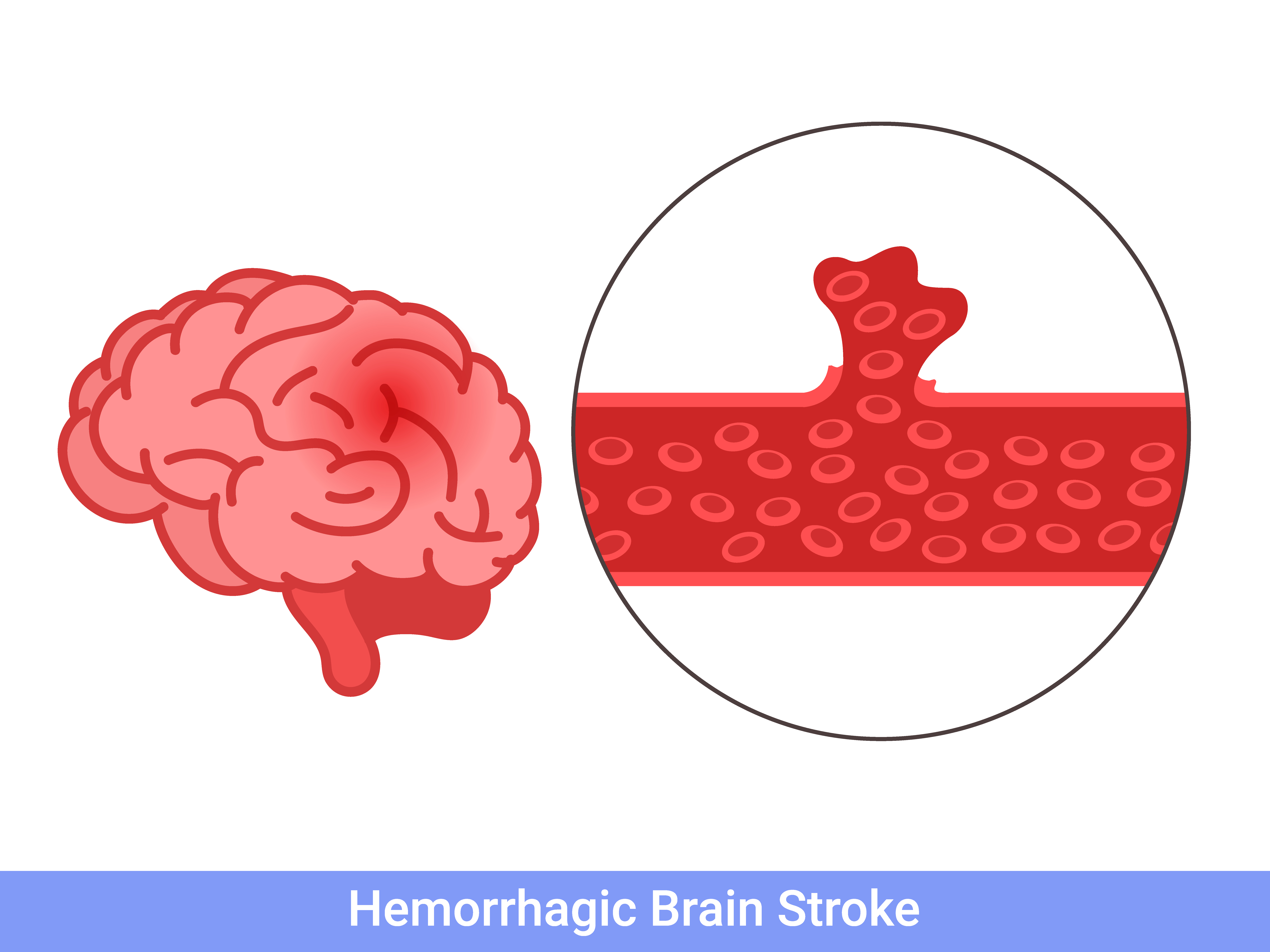
A brain aneurysm is a weak or bulging area in the wall of a blood vessel in the brain. In many cases, small aneurysms may not cause noticeable symptoms. However, if the aneurysm grows larger or ruptures, it can lead to serious neurological emergencies, including bleeding in the brain.
Brain aneurysm symptoms and treatment depend on the size, location, and condition of the aneurysm. Early diagnosis is important for reducing risks and improving outcomes.
Can Headaches Be a Warning Sign?
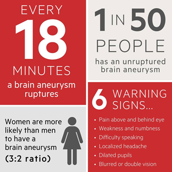
Not every headache is linked to a brain aneurysm, but certain types of headaches should never be ignored. One of the most concerning symptoms is a sudden and extremely severe headache, often described as the “worst headache of life.” This type of headache is sometimes called a thunderclap headache and may occur when an aneurysm leaks or ruptures. According to Dr. Vivek Gupta, sudden severe headaches accompanied by neurological symptoms should be evaluated immediately to rule out serious conditions such as aneurysm rupture or bleeding in the brain.
Other Symptoms of Brain Aneurysm
In addition to headaches, several other neurological emergency symptoms may appear depending on the aneurysm’s size or whether it has ruptured.
- Sudden Severe Headache – A rapid and intense headache that develops without warning can be a major red flag.
- Vision Problems – Blurred or double vision may occur if the aneurysm presses on nearby nerves.
- Neck Pain or Stiffness – Some individuals may experience stiffness or pain around the neck area.
- Nausea and Vomiting – These symptoms may accompany sudden pressure changes in the brain.
- Sensitivity to Light – Increased sensitivity to light may occur during aneurysm-related headaches.
- Weakness or Difficulty Speaking – Neurological symptoms such as facial weakness, confusion, or trouble speaking require urgent medical attention.
When Should You Seek Medical Help?
While most headaches are not dangerous, certain warning signs require immediate evaluation. Seek medical attention if headaches are:
- Sudden and extremely severe
- Different from usual headache patterns
- Accompanied by vomiting or confusion
- Associated with vision changes or weakness
- Occurring after loss of consciousness
Prompt diagnosis through brain imaging and neurological assessment can help identify potential aneurysms early.
Reducing Brain Health Risks
Maintaining healthy blood pressure, avoiding smoking, managing stress, and scheduling regular health check-ups may help support vascular and neurological health. Individuals with a family history of aneurysms or stroke should be especially mindful of persistent or unusual headache symptoms.
Awareness of headache warning signs and early medical evaluation can make a significant difference in preventing serious neurological complications. Understanding the connection between severe headaches and brain aneurysm risk is an important step toward protecting long-term brain health. Consult us for a timely brain health evaluation and advanced neurological care if you experience persistent or sudden severe headaches.