Recovering after brain aneurysm treatment is a crucial phase that significantly influences long-term brain health and overall quality of life. Whether the aneurysm was treated through surgical clipping or an endovascular procedure such as coiling, the brain needs time, protection, and structured care to heal effectively. As emphasized by Dr. Vivek Gupta, understanding the right practices to follow—and habits to avoid—can help patients recover safely and reduce the risk of complications.
Why Post-Treatment Care Matters After a Brain Aneurysm
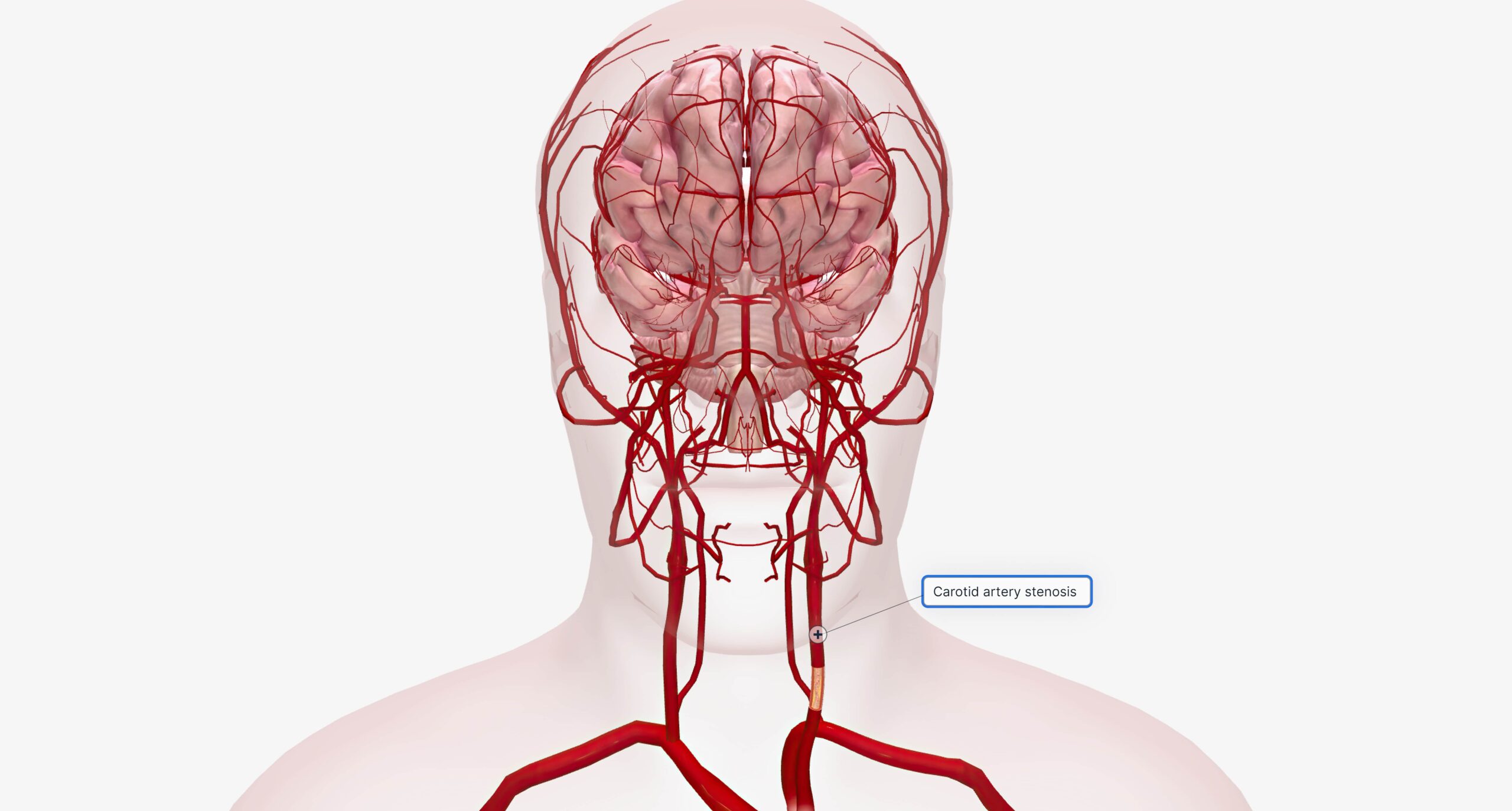
Brain aneurysm treatment addresses the immediate risk of rupture, but recovery continues long after the procedure. The brain is sensitive during the healing phase, and even minor stressors can impact recovery. Proper post-aneurysm care helps prevent neurological complications, supports cognitive recovery, and promotes long-term brain stability.
Do’s After Brain Aneurysm Treatment
Following recommended care guidelines can make a meaningful difference in recovery.
1. Attend All Follow-Up Appointments
Regular follow-ups allow doctors to monitor healing, assess neurological function, and ensure the treated aneurysm remains stable.
2. Take Prescribed Medications Consistently
Medications may be given to manage blood pressure, prevent seizures, or control pain. Following the ns is essential for brain recovery.
3. Prioritize Rest and Sleep
Adequate rest supports brain healing and reduces fatigue, headaches, and cognitive strain.
4. Resume Activities Gradually
Light daily activities can be reintroduced slowly, based on medical advice, to avoid overloading the brain during recovery.
5. Maintain a Balanced, Nutritious Diet
Proper nutrition supports brain function, energy levels, and overall healing.
Don’ts After Brain Aneurysm Treatment
Certain activities and habits can interfere with recovery and should be avoided.
1. Do Not Ignore New or Worsening Symptoms
Severe headaches, vision problems, confusion, weakness, or seizures require immediate medical attention.
2. Avoid Heavy Physical Strain
Strenuous exercise, heavy lifting, or sudden exertion can increase pressure and should be avoided until cleared by a doctor.
3. Do Not Skip Medications or Follow-Ups
Missing medications or appointments may increase the risk of complications or delayed recovery.
4. Avoid Smoking and Alcohol
Both can negatively affect brain healing and increase the risk of future neurological problems.
Long-Term Brain Health After Aneurysm Treatment
Recovery does not end once the initial healing phase is over. Long-term brain health depends on consistent follow-up care, stress management, cognitive rest, and lifestyle adjustments. Emotional well-being is also important, as anxiety or mood changes can occur after brain aneurysm treatment. Seeking support when needed can help improve overall recovery and confidence.
Every patient’s recovery timeline is different, but informed care choices play a powerful role in long-term outcomes.
Recovering after a brain aneurysm? Please consult us for expert post-treatment care and personalized guidance to support safe recovery and long-term brain health.